
Human TIGIT ELISA Kit | EL-213-20
Cat. No: EL-213-20
$859.99 USD
96 wells
| Target protein | TIGIT |
|---|---|
| Gene ID | 201633 |
| Reactivity | Human |
| Detection range | 0.32-20 ng/mL |
| Sample type | serum, plasma, tissue homogenates and other biological fluids |
| Sample volume | 100 µL |
| Assay time | 3h 20min |
| Precision | Intra-assay CV <8%. Inter-assay CV <10% |
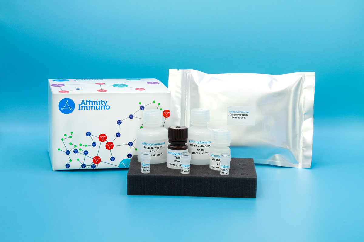
| Components | Pre-Coated Microplate - 12 strips x 8 wells (4°C/-20°C) Standard (Lyophilized) - 2 vials (4°C/-20°C) Biotinylated Antibody (100×) - 120 μL (4°C/-20°C) Streptavidin-HRP (100×) - 120 μL (4°C/-20°C) Standard/Sample Diluent Buffer - 20 mL (4°C/-20°C) Biotinylated Antibody Diluent - 12 mL (4°C/-20°C) HRP Diluent - 12 mL (4°C/-20°C) Wash Buffer (25×) - 20 mL (4°C/-20°C) TMB Substrate Solution - 10 mL (4°C/-20°C (store in dark)) Stop Reagent - 6 mL (4°C/-20°C) Plate Covers - 2 pieces (RT) |
| Storage | 4°C/-20°C |
Introduction
TIGIT is an inhibitory immune checkpoint receptor found on activated and memory T cells, regulatory T cells, and NK cells, built from a single IgV extracellular domain, a transmembrane segment, and a cytoplasmic tail carrying an ITIM motif. Its main ligand, CD155 (PVR), is displayed at high levels on dendritic cells, fibroblasts, and endothelial cells, while related proteins CD112 and CD113 bind with lower affinity; engagement of any of these ligands triggers phosphorylation of the TIGIT tail and recruitment of adaptor and phosphatase proteins that dampen PI3K, MAPK, and NF-kB signaling inside the lymphocyte. The functional outcome is a shift toward an anti-inflammatory cytokine profile, with reduced IL-12 and increased IL-10 output, along with promotion of tolerogenic dendritic cell maturation and progressive functional exhaustion of chronically stimulated T cells, including tumor-infiltrating lymphocytes. Because this same pathway also helps coordinate follicular helper T cell interactions with dendritic cells during antibody responses, TIGIT sits at the intersection of tumor immune evasion and normal regulation of T-cell-dependent B-cell help, making it an actively pursued checkpoint-blockade target.
Because TIGIT blockade is being pursued alongside PD-1/PD-L1 inhibition in solid-tumor immunotherapy trials, soluble TIGIT is used as a pharmacodynamic and immune-monitoring biomarker in combination-therapy studies, and TIGIT signaling through CD155/PVR and Nectin-2/CD112 on dendritic and endothelial cells makes it relevant to broader immune-exhaustion research as well.
Principle of the Assay
This kit uses a sandwich enzyme immunoassay format to detect and quantify Human TIGIT. Microtiter plate wells are pre-coated with a TIGIT-specific capture antibody that immobilizes the target antigen present in standards or samples added to each well. A biotin-conjugated detection antibody, also specific to Human TIGIT, is then introduced and binds a second epitope on the captured antigen. Avidin conjugated to horseradish peroxidase (HRP) is subsequently added and binds the biotin label, completing the detection complex. Upon addition of TMB substrate solution, a colorimetric signal develops only in wells containing Human TIGIT, the biotin-conjugated detection antibody, and the avidin-HRP conjugate. The enzymatic reaction is stopped by addition of a sulphuric acid stop solution, and the resulting color change is measured spectrophotometrically at 450 nm (± 10 nm). Human TIGIT concentrations in the samples are then determined by comparing sample optical density values against a standard curve.