
Human SIRPB1 ELISA Kit | EL-213-1991801
Cat. No: EL-213-1991801
$859.99 USD
96 wells
| Target protein | SIRPB1 |
|---|---|
| Gene ID | 10326 |
| Reactivity | Human |
| Detection range | 0.32-20 ng/mL |
| Sample type | Tissue homogenates, cell lysates and other biological fluids. |
| Sample volume | 100 µL |
| Assay time | 3h 20min |
| Precision | Intra-assay CV <8%. Inter-assay CV <10% |
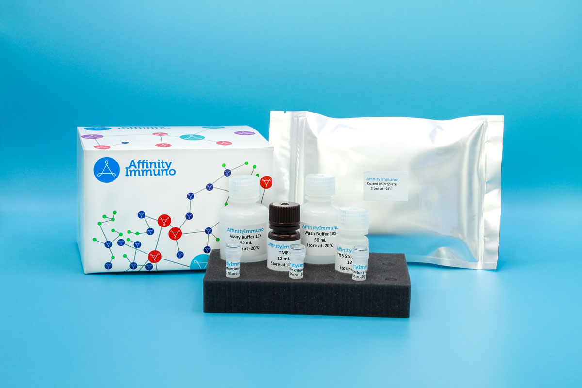
| Components | Pre-Coated Microplate - 12 strips x 8 wells (4°C/-20°C) Standard (Lyophilized) - 2 vials (4°C/-20°C) Biotinylated Antibody (100×) - 120 μL (4°C/-20°C) Streptavidin-HRP (100×) - 120 μL (4°C/-20°C) Standard/Sample Diluent Buffer - 20 mL (4°C/-20°C) Biotinylated Antibody Diluent - 12 mL (4°C/-20°C) HRP Diluent - 12 mL (4°C/-20°C) Wash Buffer (25×) - 20 mL (4°C/-20°C) TMB Substrate Solution - 10 mL (4°C/-20°C (store in dark)) Stop Reagent - 6 mL (4°C/-20°C) Plate Covers - 2 pieces (RT) |
| Storage | 4°C/-20°C |
Introduction
SIRPB1 encodes a cell-surface receptor of the signal-regulatory protein (SIRP) family within the immunoglobulin superfamily, expressed mainly on myeloid cells such as monocytes and macrophages. Unlike its inhibitory relative SIRP-alpha, SIRPB1 lacks the cytoplasmic motifs needed to dampen signaling and instead pairs with the ITAM-bearing adaptor TYROBP/DAP12 to transmit activating signals into the cell. This partnership recruits the kinase SYK and triggers downstream calcium flux along with MAPK and NF-kB pathway activity, driving myeloid cell activation rather than suppressing it. Elevated SIRPB1 expression has also been observed in glioma tissue, where it correlates with an altered immune microenvironment and worse patient outcomes.
Unlike its inhibitory relative SIRP-alpha, SIRPB1 activates rather than restrains myeloid cells, and its elevated expression in glioma tissue correlating with an altered immune microenvironment and worse patient outcomes makes this assay relevant to neuro-oncology tumor-immune-microenvironment research specifically.
Principle of the Assay
This kit uses a sandwich enzyme immunoassay format to detect Human SIRPB1. Microtiter plate wells are pre-coated with a SIRPB1-specific capture antibody, which binds the target when standards or samples are introduced. A biotin-conjugated detection antibody specific to Human SIRPB1 is then added and binds a second epitope on the captured analyte. Avidin-conjugated horseradish peroxidase (HRP) is subsequently added to each well, linking enzymatic activity to the captured complex via the biotin–avidin interaction. Upon addition of TMB substrate, a colorimetric signal develops exclusively in wells containing Human SIRPB1, biotin-conjugated antibody, and Avidin-HRP. The enzymatic reaction is stopped by addition of a sulphuric acid solution, and absorbance is measured at 450 nm (± 10 nm). Sample concentrations are determined by comparing sample optical density values against a standard curve.