
Human ROR1 ELISA Kit | EL-213-18151801
Cat. No: EL-213-18151801
$859.99 USD
96 wells
| Target protein | ROR1 |
|---|---|
| Gene ID | 4919 |
| Reactivity | Human |
| Detection range | 0.16-10 ng/mL |
| Sample type | Tissue homogenates, cell lysates and other biological fluids |
| Sample volume | 100 µL |
| Assay time | 3h 20min |
| Precision | Intra-assay CV <8%. Inter-assay CV <10% |
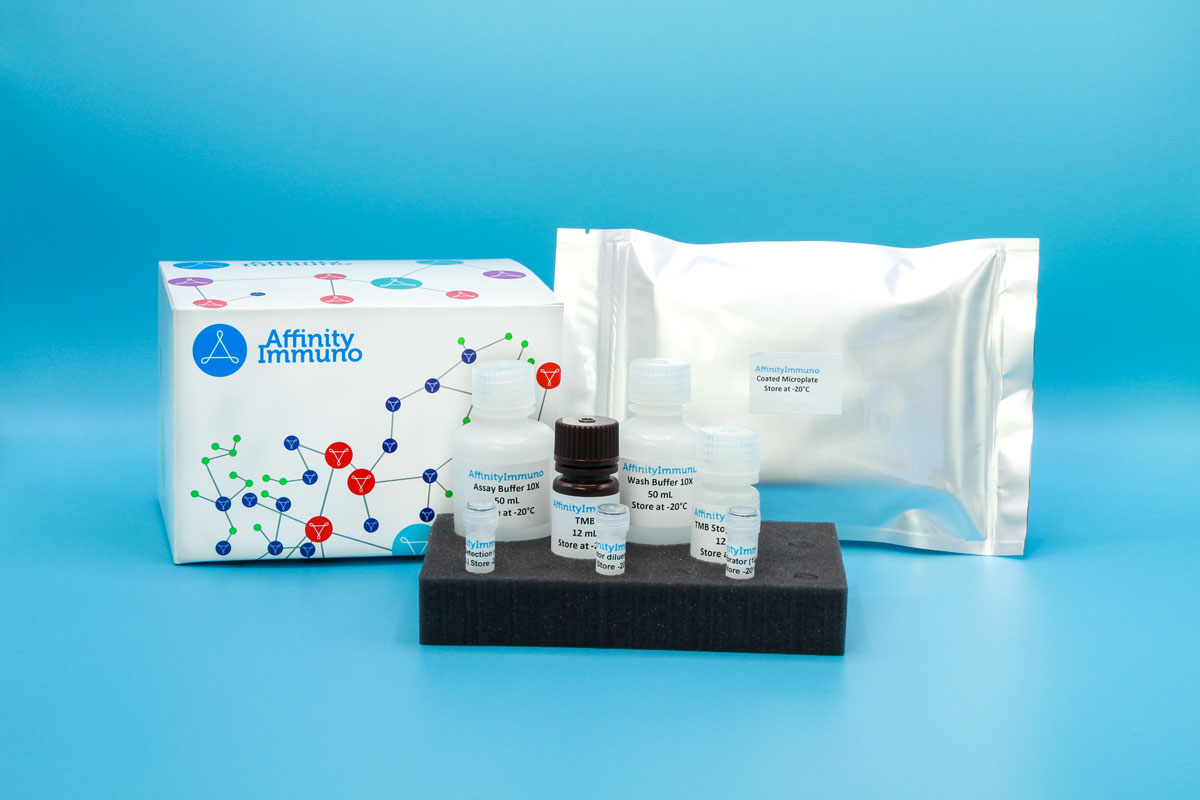
| Components | Pre-Coated Microplate - 12 strips x 8 wells (4°C/-20°C) Standard (Lyophilized) - 2 vials (4°C/-20°C) Biotinylated Antibody (100×) - 120 μL (4°C/-20°C) Streptavidin-HRP (100×) - 120 μL (4°C/-20°C) Standard/Sample Diluent Buffer - 20 mL (4°C/-20°C) Biotinylated Antibody Diluent - 12 mL (4°C/-20°C) HRP Diluent - 12 mL (4°C/-20°C) Wash Buffer (25×) - 20 mL (4°C/-20°C) TMB Substrate Solution - 10 mL (4°C/-20°C (store in dark)) Stop Reagent - 6 mL (4°C/-20°C) Plate Covers - 2 pieces (RT) |
| Storage | 4°C/-20°C |
Introduction
ROR1 encodes a type I transmembrane glycoprotein whose extracellular region combines kringle, frizzled-related, cysteine-rich, and immunoglobulin-like domains, giving it the overall architecture of a receptor tyrosine kinase, yet its intracellular kinase domain is catalytically dead and does not appear to function as a true enzyme in cells. Instead, ROR1 works as a receptor for the non-canonical Wnt ligand WNT5A, triggering NF-kB signaling along with planar-cell-polarity and calcium-dependent pathways that influence cell division, migration, and chemotaxis, while apparently also dampening signaling from the canonical ligand WNT3A. The receptor is abundant during early embryonic development, including a specific requirement for spiral ganglion neurons to properly innervate auditory hair cells in the inner ear, but its expression falls to very low levels in most healthy adult tissues. That embryonic pattern re-emerges in a wide range of malignancies, including chronic lymphocytic leukemia, mantle cell lymphoma, and cancers of the ovary, breast, prostate, lung, and colon, where ROR1 partners with IGFBP5 and ERBB2 to activate CREB-driven survival signaling and support tumor cell growth.
ROR1’s re-emergence in chronic lymphocytic leukemia, mantle cell lymphoma, and several solid tumors — while remaining nearly absent from healthy adult tissue — has driven an active antibody-therapy pipeline (cirmtuzumab/zilovertamab) aimed at blocking its WNT5A-driven survival signaling, giving this assay direct relevance to hematologic malignancy and leukemia-stem-cell research.
Principle of the Assay
This kit utilizes a sandwich enzyme immunoassay format. The microtiter plate wells are pre-coated with a capture antibody specific to Human ROR1, which binds the target analyte present in standards or samples added during the first incubation step. A biotin-conjugated detection antibody, also specific to Human ROR1, is then introduced and binds a second epitope on the captured analyte. Avidin conjugated to horseradish peroxidase (HRP) is subsequently added to each well, linking to the biotin-conjugated detection antibody. Upon addition of TMB substrate solution, a colorimetric signal develops only in wells where Human ROR1, the biotin-conjugated antibody, and avidin-HRP are all present. The enzymatic reaction is terminated by the addition of a sulphuric acid stop solution, and absorbance is measured spectrophotometrically at 450 nm (± 10 nm). Sample concentrations are determined by comparing measured optical density values against a standard curve.