
Human Irisin ELISA Kit | EL-213-09
Cat. No: EL-213-09
$859.99 USD
96 wells
| Target protein | Irisin |
|---|---|
| Alternative name | FNDC5 |
| Gene ID | 252995 |
| Reactivity | Human |
| Detection range | 0.32-20 ng/mL |
| Sample type | serum, plasma, tissue homogenates and other biological fluids |
| Sample volume | 100 µL |
| Assay time | 3h 20min |
| Precision | Intra-assay CV <8%. Inter-assay CV <10% |
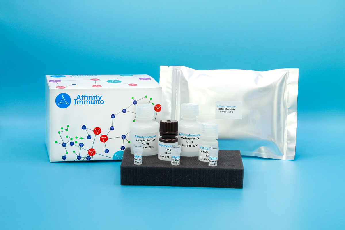
| Components | Pre-Coated Microplate - 12 strips x 8 wells (4°C/-20°C) Standard (Lyophilized) - 2 vials (4°C/-20°C) Biotinylated Antibody (100×) - 120 μL (4°C/-20°C) Streptavidin-HRP (100×) - 120 μL (4°C/-20°C) Standard/Sample Diluent Buffer - 20 mL (4°C/-20°C) Biotinylated Antibody Diluent - 12 mL (4°C/-20°C) HRP Diluent - 12 mL (4°C/-20°C) Wash Buffer (25×) - 20 mL (4°C/-20°C) TMB Substrate Solution - 10 mL (4°C/-20°C (store in dark)) Stop Reagent - 6 mL (4°C/-20°C) Plate Covers - 2 pieces (RT) |
| Storage | 4°C/-20°C |
Introduction
Irisin is the circulating hormone fragment released from FNDC5, a muscle-membrane protein initially made as a roughly 212-residue precursor with an extracellular fibronectin type III domain, a single transmembrane segment, and a short cytoplasmic tail. Physical exertion drives proteolytic shedding of the extracellular portion into the bloodstream as a glycosylated, approximately 12 kDa peptide, giving irisin its identity as an exercise-induced myokine. Once released, it acts on adipose tissue to promote a shift from white to brown-like fat by raising UCP1 levels, partly through PPARA signaling, and irisin-linked MAPK activity has separately been tied to bone formation and neuronal differentiation. FNDC5 transcripts undergo alternative splicing, and translation of the human precursor unusually begins from a non-AUG start codon that functions as an ordinary AUG start site in other species.
Irisin remains one of the most actively studied exercise biomarkers for its proposed white-to-brown fat conversion and bone-formation effects, and is frequently paired with adropin in metabolic research panels — though a well-known 2015 Scientific Reports paper raised antibody-specificity concerns about early commercial irisin ELISAs, so featuring your kit’s own specificity/validation data prominently would directly address a question researchers in this field are primed to ask.
Principle of the Assay
This kit utilizes a sandwich enzyme immunoassay format in which the microtiter plate wells are pre-coated with a capture antibody specific to Human Irisin. Standards or samples are pipetted into the appropriate wells alongside a biotin-conjugated detection antibody that also recognizes Human Irisin. Avidin conjugated to horseradish peroxidase (HRP) is then added to each well and allowed to incubate, linking enzymatic activity to any captured analyte via the biotin–avidin interaction. Upon addition of TMB substrate solution, a colorimetric signal develops exclusively in wells where Human Irisin, the biotin-conjugated antibody, and the HRP-conjugated avidin are all present. The enzymatic reaction is stopped by the addition of a sulphuric acid solution, and absorbance is read spectrophotometrically at 450 nm (± 10 nm). Human Irisin concentrations in the samples are determined by interpolating the measured optical densities against a standard curve.