
Human IL36RN ELISA Kit | EL-213-91236
Cat. No: EL-213-91236
$859.99 USD
96 wells
| Target protein | IL36RN |
|---|---|
| Gene ID | 26525 |
| Reactivity | Human |
| Detection range | 15.63-1000 pg/mL |
| Sample type | Serum, plasma, tissue homogenates, cell lysates, cell culture supernates and other biological fluids |
| Sample volume | 100 µL |
| Assay time | 3h 20min |
| Precision | Intra-assay CV <8%. Inter-assay CV <10% |
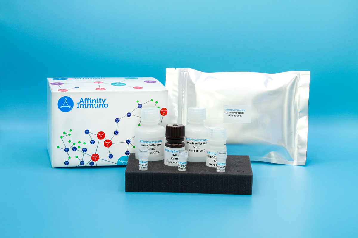
| Components | Pre-Coated Microplate - 12 strips x 8 wells (4°C/-20°C) Standard (Lyophilized) - 2 vials (4°C/-20°C) Biotinylated Antibody (100×) - 120 μL (4°C/-20°C) Streptavidin-HRP (100×) - 120 μL (4°C/-20°C) Standard/Sample Diluent Buffer - 20 mL (4°C/-20°C) Biotinylated Antibody Diluent - 12 mL (4°C/-20°C) HRP Diluent - 12 mL (4°C/-20°C) Wash Buffer (25×) - 20 mL (4°C/-20°C) TMB Substrate Solution - 10 mL (4°C/-20°C (store in dark)) Stop Reagent - 6 mL (4°C/-20°C) Plate Covers - 2 pieces (RT) |
| Storage | 4°C/-20°C |
Introduction
IL36RN encodes the interleukin-36 receptor antagonist, a secreted regulator that keeps inflammatory signaling in check at epithelial surfaces such as the skin. It works by occupying the IL-36 receptor chain (IL1RL2) so that the pro-inflammatory cytokines IL-36 alpha, beta, and gamma cannot dock there and recruit their signaling co-receptor, effectively muting a pathway that closely parallels classical IL-1 biology. This antagonist activity is thought to help dampen local innate immune responses, including reactions to fungal exposure, and may also engage an anti-inflammatory adaptor protein to reinforce the brake it puts on IL-36 signaling. Loss-of-function changes in this gene remove that brake and are the recognized cause of a severe, flare-prone pustular skin disease, underscoring how tightly IL-36 activity must be controlled for normal epidermal homeostasis.
Because loss-of-function IL36RN mutations are the recognized genetic cause of generalized pustular psoriasis — the same disease now treatable with the IL-36-receptor-blocking antibody spesolimab — this assay ties together genetic, biomarker, and drug-development research on the same pathway as our IL-36alpha and IL-36beta kits.
Principle of the Assay
This sandwich enzyme immunoassay kit uses a microtiter plate pre-coated with a capture antibody specific to Human IL36RN (FIL1δ). During incubation, IL36RN present in standards or samples is bound by the capture antibody, after which a biotin-conjugated detection antibody targeting a second epitope on Human IL36RN is added to each well. Avidin conjugated to horseradish peroxidase (HRP) is then introduced and binds the biotin-conjugated detection antibody. Upon addition of TMB substrate, a colorimetric signal develops exclusively in wells where Human IL36RN, the biotin-conjugated antibody, and avidin-HRP are all present. The enzymatic reaction is stopped by the addition of a sulphuric acid solution, and absorbance is measured spectrophotometrically at 450 nm (± 10 nm). Sample concentrations are determined by comparing sample optical density values against a standard curve.