
Human GFAP ELISA Kit | EL-213-073
Cat. No: EL-213-073
$859.99 USD
96 wells
| Target protein | GFAP |
|---|---|
| Gene ID | 2670 |
| Reactivity | Human |
| Detection range | 0.16-10 ng/mL |
| Sample type | serum, plasma, tissue homogenates, cell lysates, cell culture supernates and other biological fluids |
| Sample volume | 100 µL |
| Assay time | 3h 20min |
| Precision | Intra-assay CV <8%. Inter-assay CV <10% |
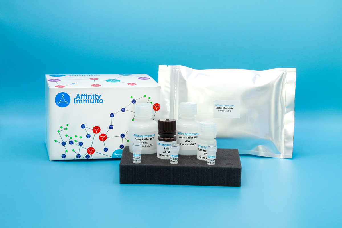
| Components | Pre-Coated Microplate - 12 strips x 8 wells (4°C/-20°C) Standard (Lyophilized) - 2 vials (4°C/-20°C) Biotinylated Antibody (100×) - 120 μL (4°C/-20°C) Streptavidin-HRP (100×) - 120 μL (4°C/-20°C) Standard/Sample Diluent Buffer - 20 mL (4°C/-20°C) Biotinylated Antibody Diluent - 12 mL (4°C/-20°C) HRP Diluent - 12 mL (4°C/-20°C) Wash Buffer (25×) - 20 mL (4°C/-20°C) TMB Substrate Solution - 10 mL (4°C/-20°C (store in dark)) Stop Reagent - 6 mL (4°C/-20°C) Plate Covers - 2 pieces (RT) |
| Storage | 4°C/-20°C |
Introduction
GFAP is a roughly 50-kilodalton intermediate filament protein that forms part of the cytoskeleton in astrocytes and remains the most reliable marker for identifying cells of astrocytic lineage within the central nervous system. Beyond structural support, it helps organize astrocyte shape and cell-adhesion contacts and contributes to maintaining the stability of myelinated white matter, with expression changing during processes like synaptic remodeling and reactive gliosis after injury. Germline mutations in the gene cause Alexander disease, a leukodystrophy that in its infantile form brings seizures, an enlarged head, and developmental regression, while later-onset forms present more gradually with ataxia and brainstem-related symptoms; the hallmark pathological finding is the astrocytic Rosenthal fiber. Because astrocyte injury releases GFAP into biofluids, measuring it in blood or cerebrospinal fluid has become a way to gauge astrocyte activation and damage, including after traumatic brain injury.
Rising GFAP levels following brain injury have made it one of the most closely watched blood-based biomarkers in traumatic brain injury and stroke research, and it remains central to studying astrogliosis in neurodegenerative disease, epilepsy, and the rare pediatric disorder Alexander disease.
Principle of the Assay
This kit uses a sandwich enzyme immunoassay format in which the microtiter plate wells are pre-coated with a capture antibody specific to Human GFAP. Standards or samples are added to the appropriate wells together with a biotin-conjugated detection antibody that recognizes a second epitope on Human GFAP. Avidin conjugated to horseradish peroxidase (HRP) is then added to each well and allowed to incubate, linking enzymatic activity to any captured analyte. Upon addition of TMB substrate solution, a colorimetric signal develops exclusively in wells containing Human GFAP, the biotin-conjugated detection antibody, and avidin-HRP. The enzymatic reaction is stopped by the addition of a sulphuric acid solution, and absorbance is measured spectrophotometrically at 450 nm ± 10 nm. Sample concentrations are determined by comparing the optical density of each sample against a standard curve.