
Human EpCAM ELISA Kit | EL-213-05
Cat. No: EL-213-05
$859.99 USD
96 wells
| Target protein | EpCAM |
|---|---|
| Gene ID | 4072 |
| Reactivity | Human |
| Detection range | 31.25-2000 pg/mL |
| Sample type | Serum, plasma, tissue homogenates, cell lysates and other biological fluids. |
| Sample volume | 100 µL |
| Assay time | 3h 20min |
| Precision | Intra-assay CV <8%. Inter-assay CV <10% |
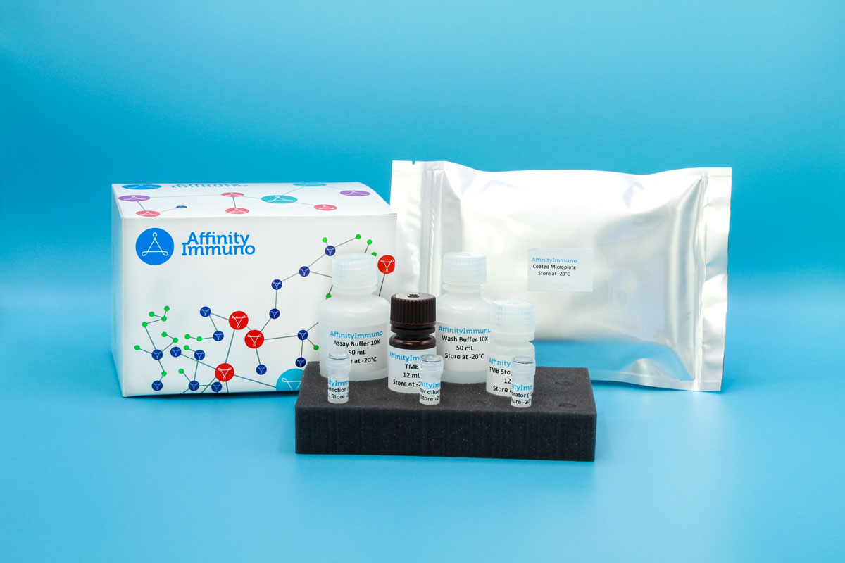
| Components | Pre-Coated Microplate - 12 strips x 8 wells (4°C/-20°C) Standard (Lyophilized) - 2 vials (4°C/-20°C) Biotinylated Antibody (100×) - 120 μL (4°C/-20°C) Streptavidin-HRP (100×) - 120 μL (4°C/-20°C) Standard/Sample Diluent Buffer - 20 mL (4°C/-20°C) Biotinylated Antibody Diluent - 12 mL (4°C/-20°C) HRP Diluent - 12 mL (4°C/-20°C) Wash Buffer (25×) - 20 mL (4°C/-20°C) TMB Substrate Solution - 10 mL (4°C/-20°C (store in dark)) Stop Reagent - 6 mL (4°C/-20°C) Plate Covers - 2 pieces (RT) |
| Storage | 4°C/-20°C |
Introduction
EPCAM encodes a small type I transmembrane glycoprotein, also known as TROP-1 or CD326, that sits on the surface of most epithelial cells and mediates calcium-independent, homotypic contacts between neighboring cells, including a direct physical connection between the epithelial cells lining the gut and the immune cells stationed within that lining, known as intraepithelial lymphocytes. Beyond simple adhesion, its intracellular fragment can be released by regulated proteolysis and travel to the nucleus, where it helps drive expression of growth-related genes such as MYC and cyclins, supporting proliferation of embryonic and tumor-initiating stem-like cells. Because this protein is displayed at high density on gastrointestinal and other carcinomas but is comparatively restricted on healthy tissue, it has become a favored target for antibody-based cancer immunotherapies. Loss-of-function mutations instead cause a severe congenital diarrheal disorder marked by intestinal villous atrophy and epithelial tufting, and EPCAM deletions are also a recognized, less common cause of hereditary colorectal cancer predisposition through silencing of the neighboring mismatch-repair gene MSH2.
EpCAM’s high, selective density on gastrointestinal and other carcinomas has made it both a long-standing antibody-therapy target and the molecular basis for widely used circulating-tumor-cell capture technologies, giving this assay relevance to liquid-biopsy and cancer-diagnostics research in addition to its established role in hereditary colorectal cancer studies.
Principle of the Assay
This kit utilizes a sandwich enzyme immunoassay format in which the microtiter plate wells are pre-coated with a capture antibody specific to human EpCAM. Standards or samples are pipetted into the appropriate wells together with a biotin-conjugated detection antibody that also recognizes human EpCAM. Avidin conjugated to horseradish peroxidase (HRP) is then added to each well and allowed to incubate, linking enzymatic activity to any captured analyte. Upon addition of TMB substrate solution, a colorimetric signal develops exclusively in wells containing human EpCAM, the biotin-conjugated detection antibody, and Avidin-HRP. The enzymatic reaction is stopped by the addition of sulphuric acid solution, and the resulting color change is measured spectrophotometrically at 450 nm (± 10 nm). Human EpCAM concentrations in the samples are subsequently calculated by comparing sample optical density values against a standard curve.