
Human DLL3 ELISA Kit | EL-213-4121203
Cat. No: EL-213-4121203
$859.99 USD
96 wells
| Target protein | DLL3 |
|---|---|
| Gene ID | 10683 |
| Reactivity | Human |
| Detection range | 0.16-10 ng/mL |
| Sample type | Tissue homogenates and other biological fluids. |
| Sample volume | 100 µL |
| Assay time | 3h 20min |
| Precision | Intra-assay CV <8%. Inter-assay CV <10% |
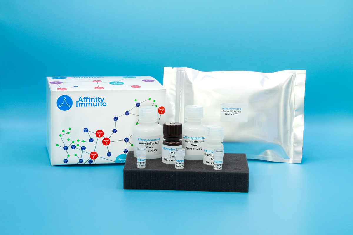
| Components | Pre-Coated Microplate - 12 strips x 8 wells (4°C/-20°C) Standard (Lyophilized) - 2 vials (4°C/-20°C) Biotinylated Antibody (100×) - 120 μL (4°C/-20°C) Streptavidin-HRP (100×) - 120 μL (4°C/-20°C) Standard/Sample Diluent Buffer - 20 mL (4°C/-20°C) Biotinylated Antibody Diluent - 12 mL (4°C/-20°C) HRP Diluent - 12 mL (4°C/-20°C) Wash Buffer (25×) - 20 mL (4°C/-20°C) TMB Substrate Solution - 10 mL (4°C/-20°C (store in dark)) Stop Reagent - 6 mL (4°C/-20°C) Plate Covers - 2 pieces (RT) |
| Storage | 4°C/-20°C |
Introduction
DLL3 belongs to the family of Delta-class ligands that normally trigger Notch receptor signaling on neighboring cells, but it behaves in an unusual, almost opposite way. Rather than sitting on the cell surface ready to activate Notch on an adjacent cell the way its relatives DLL1, DLL4, and the Jagged ligands do, DLL3 is largely retained inside the Golgi apparatus and, when it does reach the membrane, acts within the same cell to interfere with Notch signaling rather than switch it on elsewhere. This built-in brake on Notch activity helps steer developing cells toward becoming neurons instead of remaining progenitors and is required for properly dividing the embryonic body plan into segments, since loss of the gene disrupts vertebral and rib formation. That same restraining role in a pathway central to neuroendocrine cell identity likely explains why DLL3 becomes strikingly overexpressed on the surface of small cell lung cancer and other high-grade neuroendocrine tumors while remaining scarce on most normal adult tissue, a pattern that has made it an actively pursued target for antibody-based cancer therapies.
DLL3 has moved to the center of small cell lung cancer research since tarlatamab, a DLL3-targeting bispecific T-cell engager, reached approval, with recent studies exploring DLL3 expression (including in circulating tumor cells) as a predictive biomarker for response to tarlatamab and related therapies.
Principle of the Assay
This kit uses a sandwich enzyme immunoassay format in which the microtiter plate wells are pre-coated with a DLL3-specific capture antibody. Standards or samples are introduced into the appropriate wells alongside a biotin-conjugated detection antibody that also recognizes Human DLL3. Avidin conjugated to horseradish peroxidase (HRP) is then added to each well and allowed to incubate. Upon addition of TMB substrate solution, a colorimetric signal develops exclusively in wells containing Human DLL3, the biotin-conjugated detection antibody, and the avidin-HRP conjugate. The enzymatic reaction is stopped by the addition of a sulphuric acid stop solution, and the resulting color change is measured spectrophotometrically at 450 nm (± 10 nm). Sample concentrations are determined by comparing the optical density of each sample against a standard curve.