
Human CEACAM1 ELISA Kit | EL-213-35101
Cat. No: EL-213-35101
$859.99 USD
96 wells
| Target protein | CEACAM1 |
|---|---|
| Gene ID | 634 |
| Reactivity | Human |
| Detection range | 1.57-100 ng/mL |
| Assay time | 20min |
| Precision | Intra-assay CV <8%. Inter-assay CV <10% |
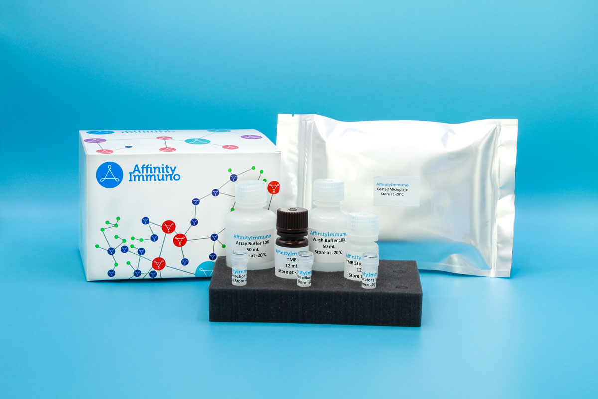
| Components | Pre-Coated Microplate - 12 strips x 8 wells (4°C/-20°C) Standard Microplate - 2 strips x 8 wells (4°C/-20°C) HRP Conjugate Antibody (100×) - 70 μL (4°C/-20°C (store in dark)) Standard/Sample Diluent Buffer - 24 mL (4°C/-20°C) HRP Conjugate Diluent - 10 mL (4°C/-20°C) Wash Buffer (25×) - 24 mL (4°C/-20°C) TMB Substrate Solution - 12 mL (4°C/-20°C (store in dark)) Stop Reagent - 7 mL (4°C/-20°C) Plate Covers - 2 pieces (4°C/-20°C) Instruction Manual - 1 (RT) |
| Storage | 4°C/-20°C |
Introduction
CEACAM1, also known as CD66a or biliary glycoprotein, is an immunoglobulin-superfamily adhesion molecule expressed on epithelial cells, endothelial cells and multiple leukocyte populations. It binds to itself and to related CEACAM-family proteins on neighboring cells, and its cytoplasmic tail carries an ITIM motif that recruits phosphatase activity to dampen signaling downstream of the T-cell receptor and NK-activating receptors. Through this mechanism it acts as a checkpoint-like inhibitory receptor that restrains cytotoxic lymphocyte activity, while separately contributing to blood vessel remodeling, insulin clearance in the liver, and tissue architecture during development. Because of these overlapping roles in immune regulation and tissue organization, CEACAM1 is studied in contexts ranging from tumor immune evasion to metabolic and vascular research.
CEACAM1’s ITIM-driven restraint of cytotoxic lymphocyte activity has made it a clinical-stage immuno-oncology target (CM24, in development by Purple Biotech) in addition to its established roles in vascular remodeling and hepatic insulin clearance, giving this assay dual relevance to checkpoint-blockade and metabolic research.
Principle of the Assay
This kit utilizes a sandwich enzyme immunoassay format. The microtiter plate is pre-coated with a capture antibody specific to Human CEACAM1; a separate set of standard plate wells pre-coated using protein-related techniques is provided alongside. Standards or samples are dispensed into the appropriate wells, followed by addition of an HRP-conjugated detection antibody specific to Human CEACAM1. Upon addition of TMB substrate solution, a colorimetric signal develops exclusively in wells containing Human CEACAM1 bound to the HRP-conjugated antibody. The enzymatic reaction is stopped by the addition of sulphuric acid solution, and absorbance is measured at 450 nm (± 10 nm). Human CEACAM1 concentrations in the samples are then calculated by comparing sample optical densities against the standard curve.