
Human CD63 ELISA Kit | EL-213-3463
Cat. No: EL-213-3463
$859.99 USD
96 wells
| Target protein | CD63 |
|---|---|
| Gene ID | 967 |
| Reactivity | Human |
| Detection range | 0.16-10 ng/mL |
| Sample type | Serum, plasma, tissue homogenates, cell lysates and other biological fluids |
| Sample volume | 100 µL |
| Assay time | 3h 20min |
| Precision | Intra-assay CV <8%. Inter-assay CV <10% |
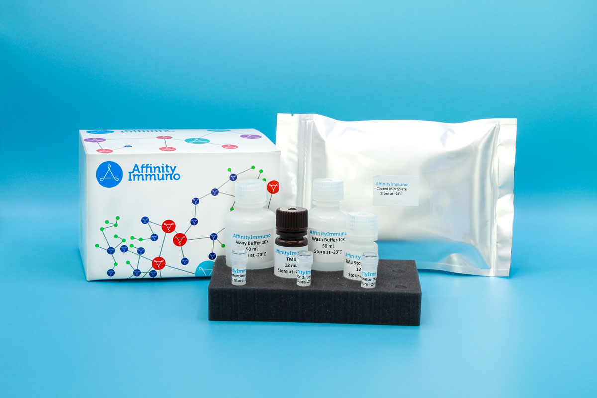
| Components | Pre-Coated Microplate - 12 strips x 8 wells (4°C/-20°C) Standard (Lyophilized) - 2 vials (4°C/-20°C) Biotinylated Antibody (100×) - 120 μL (4°C/-20°C) Streptavidin-HRP (100×) - 120 μL (4°C/-20°C) Standard/Sample Diluent Buffer - 20 mL (4°C/-20°C) Biotinylated Antibody Diluent - 12 mL (4°C/-20°C) HRP Diluent - 12 mL (4°C/-20°C) Wash Buffer (25×) - 20 mL (4°C/-20°C) TMB Substrate Solution - 10 mL (4°C/-20°C (store in dark)) Stop Reagent - 6 mL (4°C/-20°C) Plate Covers - 2 pieces (RT) |
| Storage | 4°C/-20°C |
Introduction
CD63, also designated TSPAN30, is a four-pass transmembrane glycoprotein of the tetraspanin family that resides mainly in late endosomes, multivesicular bodies and lysosomes, with a smaller pool on the plasma membrane. It serves as a docking receptor for TIMP1 and cooperates with beta-1 integrins to switch on AKT, FAK and MAP kinase signaling, effects that promote cell adhesion, spreading and migration. Its concentration in multivesicular bodies also makes it a structural marker of exosomes, and it appears on activated platelets, where it serves as an indicator of platelet activation. A deficiency in this protein produces pigment and bleeding abnormalities resembling Hermansky-Pudlak syndrome, and shifts in CD63 expression have been reported across several cancer types.
As one of the most widely used surface markers for identifying and quantifying exosomes, CD63 is central to liquid-biopsy and extracellular-vesicle research pipelines, and its appearance on activated platelets separately makes it a marker of platelet activation in hemostasis studies.
Principle of the Assay
This kit uses a sandwich enzyme immunoassay format to measure human CD63 (TSPAN30). Microtiter plate wells are pre-coated with a CD63-specific capture antibody that binds the target analyte present in standards or samples. A biotin-conjugated detection antibody, also specific to human CD63, is then added and binds a second epitope on the captured analyte. Avidin-conjugated horseradish peroxidase (HRP) is subsequently introduced and binds the biotin label. Upon addition of TMB substrate solution, a colorimetric signal develops only in wells where CD63, the biotin-conjugated detection antibody, and avidin-HRP are all present. The enzymatic reaction is stopped by the addition of sulphuric acid solution, and absorbance is measured spectrophotometrically at 450 nm ± 10 nm. CD63 concentrations in the samples are determined by comparing sample optical density values against a standard curve.