
Human CD47 ELISA Kit | EL-213-3447
Cat. No: EL-213-3447
$859.99 USD
96 wells
| Target protein | CD47 |
|---|---|
| Gene ID | 961 |
| Reactivity | Human |
| Detection range | 0.32-20 ng/mL |
| Sample type | Tissue homogenates, cell lysates and other biological fluids. |
| Sample volume | 100 µL |
| Assay time | 3h 20min |
| Precision | Intra-assay CV <8%. Inter-assay CV <10% |
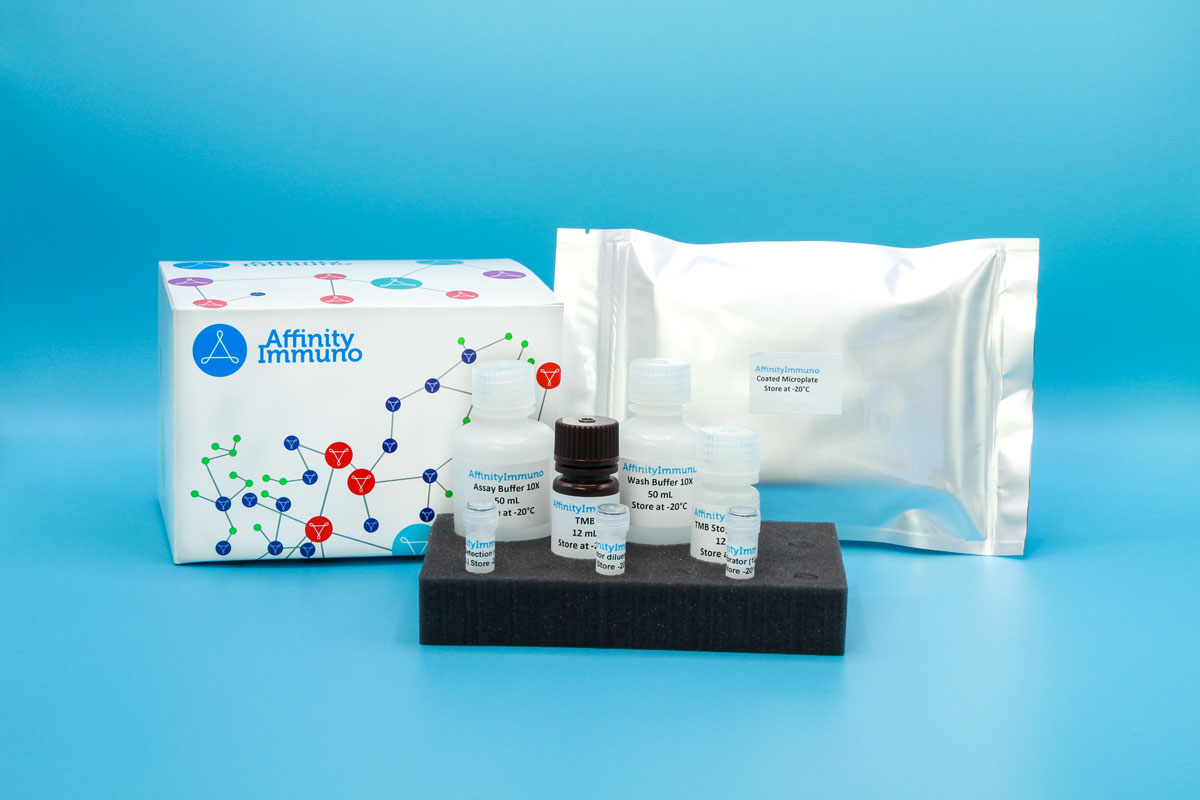
| Components | Pre-Coated Microplate - 12 strips x 8 wells (4°C/-20°C) Standard (Lyophilized) - 2 vials (4°C/-20°C) Biotinylated Antibody (100×) - 120 μL (4°C/-20°C) Streptavidin-HRP (100×) - 120 μL (4°C/-20°C) Standard/Sample Diluent Buffer - 20 mL (4°C/-20°C) Biotinylated Antibody Diluent - 12 mL (4°C/-20°C) HRP Diluent - 12 mL (4°C/-20°C) Wash Buffer (25×) - 20 mL (4°C/-20°C) TMB Substrate Solution - 10 mL (4°C/-20°C (store in dark)) Stop Reagent - 6 mL (4°C/-20°C) Plate Covers - 2 pieces (RT) |
| Storage | 4°C/-20°C |
Introduction
CD47, also known as integrin-associated protein, is a broadly expressed multi-pass membrane receptor of the immunoglobulin superfamily found on nearly all normal tissues as well as many tumor cells. It binds thrombospondin-1 and modulates integrin-linked signaling, contributing to processes as diverse as cell adhesion, platelet function, vascular tone, and hippocampal synaptic plasticity. Its best-characterized immune role is as the ligand for SIRP-alpha on macrophages and dendritic cells; this interaction delivers a ‘do-not-eat’ signal that limits phagocytosis and dampens cytokine release, a mechanism that many cancers exploit by overexpressing CD47 to escape immune clearance. Reduced CD47 density on red blood cells lacking the Rh antigen complex further points to a role for this protein in helping normal erythrocytes avoid premature removal from circulation.
Beyond its role in tumor immune evasion via SIRP-alpha, CD47 also functions as the thrombospondin-1 receptor on platelets and helps protect circulating red blood cells from premature clearance, giving this assay relevance in hematology and vascular-adhesion studies alongside CD47-blockade immuno-oncology research.
Principle of the Assay
This kit uses a sandwich enzyme immunoassay format. The microtiter plate wells are pre-coated with a CD47 (IAP)-specific capture antibody that binds the target antigen present in standards or samples. A biotin-conjugated detection antibody, also specific to CD47, is then added and binds a second epitope on the captured antigen. Avidin conjugated to horseradish peroxidase (HRP) is subsequently introduced and binds the biotin label. Upon addition of TMB substrate solution, a colorimetric signal develops exclusively in wells where CD47, the biotin-conjugated detection antibody, and avidin-HRP are all present. The enzymatic reaction is stopped by the addition of sulfuric acid stop solution, and absorbance is measured spectrophotometrically at 450 nm ± 10 nm. CD47 concentrations in the samples are then determined by referencing the sample optical density values against a standard curve.