
Human BAFF ELISA Kit | EL-213-02
Cat. No: EL-213-02
$859.99 USD
96 wells
| Target protein | BAFF |
|---|---|
| Alternative name | TNFSF13B |
| Gene ID | 10673 |
| Reactivity | Human |
| Detection range | 78.13-5000 pg/mL |
| Sample type | serum, plasma, tissue homogenates and other biological fluids |
| Sample volume | 100 µL |
| Assay time | 3h 20min |
| Precision | Intra-assay CV <8%. Inter-assay CV <10% |
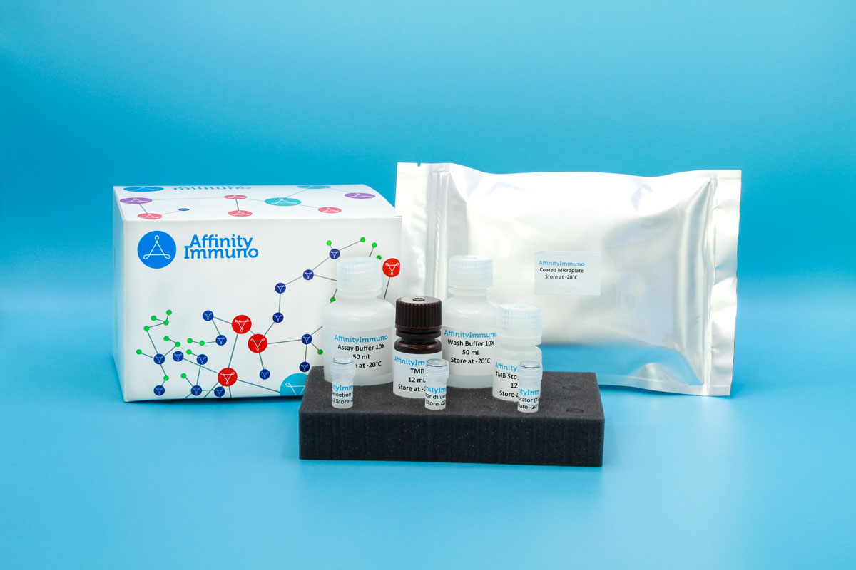
| Components | Pre-Coated Microplate - 12 strips x 8 wells (4°C/-20°C) Standard (Lyophilized) - 2 vials (4°C/-20°C) Biotinylated Antibody (100×) - 120 μL (4°C/-20°C) Streptavidin-HRP (100×) - 120 μL (4°C/-20°C) Standard/Sample Diluent Buffer - 20 mL (4°C/-20°C) Biotinylated Antibody Diluent - 12 mL (4°C/-20°C) HRP Diluent - 12 mL (4°C/-20°C) Wash Buffer (25×) - 20 mL (4°C/-20°C) TMB Substrate Solution - 10 mL (4°C/-20°C (store in dark)) Stop Reagent - 6 mL (4°C/-20°C) Plate Covers - 2 pieces (RT) |
| Storage | 4°C/-20°C |
Introduction
BAFF (also called BLyS or TNFSF13B) is a TNF-superfamily cytokine produced mainly by cells of the B-lymphocyte lineage. It signals through three distinct receptors – TACI, BCMA, and the B-cell-restricted BAFF receptor (BR3) – and, together with the related ligand APRIL, forms a dual-ligand, dual-receptor circuit that shapes humoral immunity. Through these receptors BAFF drives B-cell proliferation, differentiation, and long-term survival, while also contributing to T-cell costimulation and skewing of helper T-cell responses. Because excess BAFF activity is linked to abnormal B-cell persistence and autoimmune pathology, circulating BAFF levels are frequently monitored as a marker of B-cell-driven immune dysregulation.
Excess BAFF activity is linked to abnormal B-cell persistence and is a validated therapeutic target in systemic lupus erythematosus (the target of belimumab), so circulating BAFF is frequently monitored in autoimmune research alongside its related ligand APRIL and receptors TACI, BCMA, and BAFF-R.
Principle of the Assay
This kit uses a sandwich enzyme immunoassay format in which the microtiter plate wells are pre-coated with a capture antibody specific to Human BAFF/CD257. During incubation, any BAFF/CD257 present in standards or samples is bound by this capture antibody, and a biotin-conjugated detection antibody directed against a second epitope on Human BAFF/CD257 is added simultaneously. Avidin conjugated to horseradish peroxidase (HRP) is then introduced and binds the biotin label. Upon addition of TMB substrate solution, a colorimetric signal develops exclusively in wells containing the BAFF/CD257–biotin-antibody–avidin-HRP complex. The enzymatic reaction is stopped by the addition of sulphuric acid, and absorbance is measured at 450 nm (± 10 nm). Sample concentrations are determined by referencing the resulting optical density values against a standard curve.